
Antioxidant, Antibacterial, and Probiotic Effects of Ajuga Chamaecistus on Honeybee Enterococcus
1. Introduction
Natural products derived from medicinal plants have historically played a central role in drug discovery and the development of therapeutic agents [1-3].
Across different civilizations, plants have been used as primary sources of remedies for infectious diseases, inflammatory conditions, metabolic disorders, and other health problems [3]. In modern pharmaceutical sciences, interest in plant-derived bioactive compounds has increased significantly due to their structural diversity, biological potency, and lower toxicity compared to synthetic drugs [4]. Secondary metabolites such as phenolic acids, flavonoids, terpenoids, glycosides, and alkaloids are particularly important due to their wide range of pharmacological activities, including antioxidant, antimicrobial, anticancer, and anti-inflammatory effects [5-8]. This renewed attention has been further strengthened by the urgent need for novel therapeutic agents to address the global rise in antibiotic resistance and oxidative stress-related diseases. Oxidative stress is a biological condition characterized by an imbalance between the production of reactive oxygen species and the ability of biological systems to detoxify these reactive intermediates [9-11]. Excessive oxidative stress can lead to cellular damage through lipid peroxidation [9], protein oxidation, and Deoxyribonucleic Acid (DNA) damage, ultimately contributing to the development of chronic and degenerative diseases such as cardiovascular disorders, neurodegenerative diseases, diabetes, and cancer [10]. Antioxidants are molecules that neutralize free radicals and prevent oxidative damage. Although synthetic antioxidants have been widely used in the pharmaceutical and food industries, concerns regarding their potential adverse health effects have led researchers to explore natural alternatives [11]. Plant-based antioxidants, particularly those rich in polyphenolic compounds, exhibit strong free radical-scavenging activity and are considered safer and more effective candidates for long-term applications. In addition to oxidative stress, antimicrobial resistance has become one of the most critical global health challenges of the 21st century [12]. The overuse and misuse of antibiotics in human medicine, veterinary practice, and agriculture have accelerated the emergence of multidrug-resistant pathogens. This phenomenon has reduced the effectiveness of conventional antibiotics and increased the risk of treatment failure in infectious diseases. Consequently, there is an increasing demand for new antimicrobial agents with novel mechanisms of action. Medicinal plants represent a promising reservoir of such compounds, as many plant extracts have demonstrated inhibitory effects against a broad spectrum of bacterial pathogens [14-16]. These antimicrobial properties are often attributed to the synergistic action of multiple phytochemicals rather than a single active compound. The genus Ajuga belongs to the Lamiaceae family and comprises a large group of herbaceous plants widely distributed in temperate regions of Asia, Europe, and Africa. Species of this genus have been traditionally used in folk medicine for the treatment of various ailments, including inflammatory diseases, gastrointestinal disorders, and infections. Phytochemical studies have revealed that Ajuga species contain a variety of bioactive compounds, particularly iridoids, flavonoids, and phenolic derivatives, which are responsible for their therapeutic properties [15]. Among these species, Ajuga chamaecistus is a perennial herb native to Iran and neighboring regions. Despite its traditional medicinal use, relatively limited scientific research has been conducted on its pharmacological properties compared to other members of the genus. This lack of comprehensive investigation highlights the need for further studies to explore its potential as a source of natural antioxidants and antimicrobial agents. Probiotics have gained considerable attention in recent years due to their beneficial effects on host health, particularly on gut microbiota balance, immune system modulation, and pathogen inhibition. Probiotic microorganisms are defined as live bacteria that, when administered in adequate amounts, confer health benefits to the host [18-20]. Among them, lactic acid bacteria are the most commonly studied due to their safety and functional properties. Enterococcus durans is a member of this group and has been isolated from various natural sources, including fermented foods and insect-associated microbiota. This species has demonstrated promising probiotic characteristics, including acid and bile tolerance, antimicrobial activity against pathogenic bacteria, and potential immunomodulatory effects. These properties make it a suitable candidate for applications in functional foods and biotherapeutic formulations. Honeybees (Apis mellifera) are an important ecological and agricultural species, not only for their role in pollination but also for their complex and beneficial gut microbiota. The microbial community of honeybees plays a crucial role in their nutrition, immune defense, and overall colony health. Recent studies have shown that honeybee-associated bacteria may serve as a valuable source of novel probiotic strains with unique functional properties. These microorganisms are naturally adapted to fluctuating environmental conditions, making them potentially more resilient and effective compared to conventional probiotic strains [17]. As a result, there is growing interest in isolating and characterizing probiotic bacteria from honeybee microbiota for use in biotechnology and health-related applications. In recent years, increasing attention has been given to the interaction between plant-derived bioactive compounds and probiotic microorganisms. Plant extracts rich in polyphenols and other secondary metabolites may act as prebiotic-like substances, promoting the growth and metabolic activity of beneficial bacteria. In some cases, these compounds can enhance bacterial stress tolerance, improve cell viability, and modulate microbial metabolism. This synergistic relationship between plant extracts and probiotics offers a promising approach for developing functional food ingredients and therapeutic formulations. However, limited studies have investigated the specific effects of medicinal plant extracts on bee-derived probiotic strains, particularly in the context of Ajuga species [6-13]. Based on this background, the present study aims to investigate the antioxidant and antibacterial properties of Ajuga chamaecistus extracts and to evaluate their potential role in enhancing the growth and functional performance of Enterococcus durans isolated from honeybee Apis Mellifera Meda. The study focuses on the extraction and preliminary phytochemical evaluation of A. chamaecistus, assessment of its antioxidant and antibacterial activities, and investigation of its possible synergistic effects on probiotic viability and functionality [9]. By integrating phytochemical analysis with microbiological and probiotic evaluation, this research seeks to contribute to a better understanding of the potential applications of Iranian medicinal plants in the
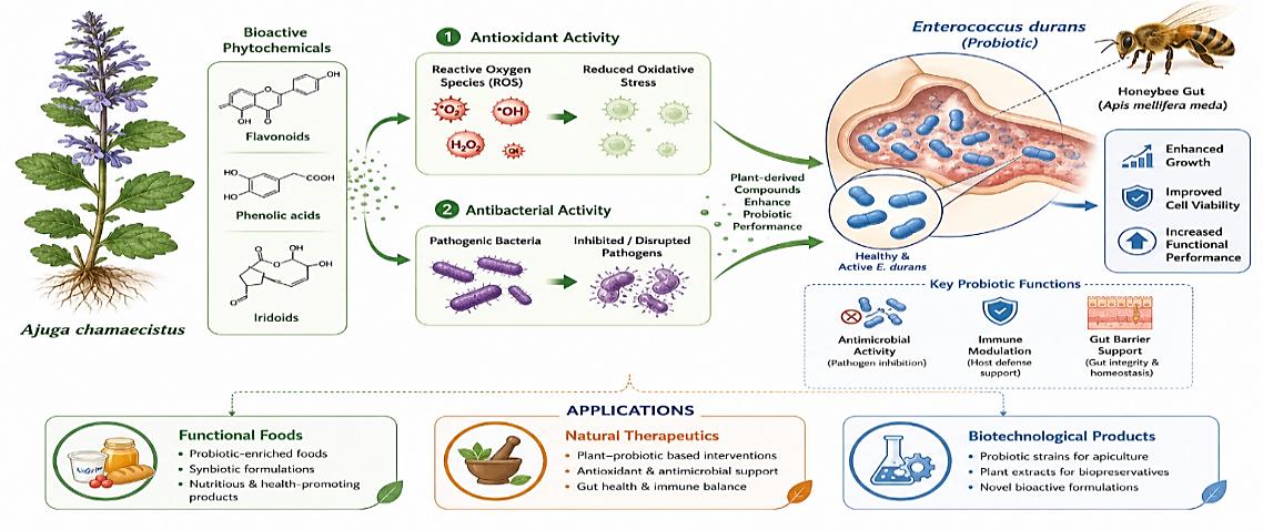
development of novel antimicrobial and probiotic-enhancing agents. The findings may also provide a scientific basis for future studies aimed at developing natural therapeutics, functional foods, and sustainable biotechnological products based on plant microbe interactions. The conceptual framework of the present study, illustrating the synergistic interaction between Ajuga chamaecistus phytochemicals and the probiotic Enterococcus durans isolated from Apis Mellifera Meda, is presented in Fig. 1.
2. Materials and Methods
2.1 Plant Material Collection and Preparation
Aerial parts of Ajuga chamaecistus were collected during the flowering stage from a natural habitat in Iran (provide exact location and date). A qualified botanist authenticated the plant material, and a voucher specimen was deposited in a recognized herbarium. The collected samples were washed with distilled water to remove impurities and shade-dried at room temperature (25–30°C) for 7–10 days. The dried material was ground into a fine powder using an electric grinder and stored in airtight containers until extraction [5], [17], [18].
2.2 Preparation of Plant Extracts
To evaluate the effect of solvent polarity on the extraction of bioactive compounds, five different solvents were used: methanol (80%), ethanol (70%), acetone (70%), ethyl acetate, and distilled water. For each extraction, 30 g of powdered plant material was mixed with 300 mL of solvent and macerated for 48 hours at room temperature with intermittent shaking. After extraction, the mixtures were filtered using Whatman No. 1 filter paper. Organic solvent extracts (methanol, ethanol, acetone, and ethyl acetate) were concentrated under reduced pressure using a rotary evaporator at 40°C. The aqueous extract was concentrated using a water bath and then freeze-dried (if available). All extracts were stored at 4°C until further analysis [18].
2.3 Determination of Total Phenolic Content
Total phenolic content of all five extracts was determined using the Folin–Ciocalteu method. Briefly, 0.5 mL of each extract (appropriately diluted) was mixed with 2.5 mL of Folin–Ciocalteu reagent (diluted 1:10 with distilled water). After 5 minutes, 2 mL of sodium carbonate solution (7.5% w/v) was added. The mixture was incubated in the dark at room temperature for 30 minutes, and absorbance was measured at 765 nm using a UV–Vis spectrophotometer. A calibration curve was prepared using gallic acid, and the results were expressed as mg Gallic Acid Equivalents (GAE) per gram of dry extract [7], [8], [19].
2.4|2,2-Diphenyl-1-Picrylhydrazyl Radical Scavenging Assay
The antioxidant activity of all five extracts was evaluated using the 2,2-Diphenyl-1-Picrylhydrazyl (DPPH) assay. A 0.1 mM DPPH solution was prepared in methanol. Different concentrations of each extract (25, 50, 100, and 200 µg/mL) were prepared. An equal volume (1 mL) of DPPH solution and extract was mixed and incubated in the dark for 30 minutes at room temperature. The absorbance was measured at 517 nm. Ascorbic acid was used as a reference standard [8]. The percentage of DPPH radical scavenging activity was calculated using the following Eq. (1):
A control −Asample
Inhibition(%) = ( ) × 100. (1)
Acontrol
2.5 Antibacterial Activity
2.5.1 Test microorganisms
The antibacterial activity of the plant extracts was evaluated against two standard pathogenic bacterial strains, Escherichia coli (gram-negative) and Staphylococcus aureus (gram-positive), as well as the probiotic strain Enterococcus durans. The reference strains of E. coli and S. aureus were obtained from a valid microbial culture collection (e.g., American Type Culture Collection (ATCC) or a university laboratory culture collection). All bacterial strains were maintained on nutrient agar slants at 4°C and subcultured before use. For experimental purposes, a loopful of each bacterial strain was inoculated into nutrient broth and incubated at 37°C for 18–24 hours to obtain fresh cultures. The bacterial suspensions were then adjusted to match the 0.5 McFarland standard, corresponding to approximately 1 × 10⁸ Colony Forming Unit (CFU)/mL. The probiotic strain Enterococcus durans, previously isolated from Apis Mellifera Meda, was cultured in De Man, Rogosa, and Sharpe (MRS) broth under the same incubation conditions. The culture was similarly adjusted to a 0.5 McFarland standard before use. All bacterial handling procedures were performed under aseptic conditions to prevent contamination [19].
2.5.2 Agar well diffusion method
Bacterial suspensions were adjusted to a 0.5 McFarland standard (~1 × 10⁸ CFU/mL). Mueller–Hinton agar plates were inoculated using sterile swabs. Wells (6 mm diameter) were made in the agar, and 50 µL of each extract at different concentrations (50, 100, and 200 mg/mL) was added. Methanol was used as a negative control, and ampicillin was used as a positive control. Plates were incubated at 37°C for 24 hours, and inhibition zones were measured in millimeters [10].
2.6 Isolation and Identification of Probiotic Strain
Worker honeybees of Apis Mellifera Meda were collected and surface-sterilized with 70% ethanol. The gut was dissected under sterile conditions and homogenized in sterile saline. Serial dilutions were prepared and plated on MRS agar. Plates were incubated at 37°C for 24–48 hours. Colonies were purified and identified as Enterococcus durans based on morphological characteristics, Gram staining, and catalase test [3].
2.7 Effect of Plant Extracts on Probiotic Growth
To evaluate the effect of different extracts on probiotic growth, MRS broth was supplemented with various concentrations of each extract (25, 50, and 100 µg/mL). The broth was inoculated with an overnight culture of Enterococcus durans and incubated at 37°C. Bacterial growth was monitored by measuring Optical Density (OD600) at 600 nm (OD600) at 0, 12, and 24 hours [14].
2.8 Statistical Analysis
All experiments were performed in triplicate, and results were expressed as mean ± standard deviation. Statistical analysis was performed using one-way ANOVA followed by appropriate post hoc tests. Differences were considered significant at p < 0.05.
3. Results Discussions
3.1 Total Phenolic Content
The total phenolic content of different extracts of Ajuga chamaecistus was determined using the Folin– Ciocalteu method and expressed as GAE. The results demonstrated a significant influence of solvent type on the extraction efficiency of phenolic compounds. Among the tested solvents, the highest phenolic content was observed in the methanolic extract, followed by ethanol, acetone, ethyl acetate, and aqueous extracts. The total phenolic content values were 95.4 ± 2.2 mg GAE/g extract for methanol, 82.7 ± 1.9 mg GAE/g extract for ethanol, 74.3 ± 2.5 mg GAE/g extract for acetone, 61.8 ± 2.1 mg GAE/g extract for ethyl acetate, and 49.6 ± 2.8 mg GAE/g extract for the aqueous extract Fig. 2. The superior performance of methanol and ethanol can be attributed to their high polarity, which enhances the solubility of phenolic compounds. In contrast, the lower phenolic yield observed in ethyl acetate and aqueous extracts may be due to their limited ability to solubilize certain classes of phenolics. These findings agree with previous studies on the Ajuga genus, which have identified solvent polarity as a key factor influencing the extraction of bioactive compounds. 3.2|2,2-Diphenyl-1-Picrylhydrazyl Radical Scavenging Activity
This figure is shown in the PDF version of the article.
The antioxidant activity of different extracts of Ajuga chamaecistus was evaluated using the DPPH radical scavenging assay. All extracts exhibited dose-dependent antioxidant activity, with increasing radical scavenging effects observed at higher concentrations (25–200 µg/mL). Among the tested extracts, the methanolic extract showed the highest antioxidant activity, followed by the ethanol, acetone, and ethyl acetate extracts, and the aqueous extract. At the highest concentration (200 µg/mL), the methanolic extract demonstrated 89.2 ± 1.7% inhibition, while ethanol, acetone, ethyl acetate, and aqueous extracts showed 81.5 ± 2.1%, 73.8 ± 2.4%, 65.2 ± 1.9%, and 58.6 ± 2.7% inhibition, respectively. The IC₅₀ values further confirmed these findings, with the methanolic extract exhibiting the lowest IC₅₀ value (42.3 µg/mL), indicating the strongest antioxidant activity. The IC₅₀ values for ethanol, acetone, ethyl acetate, and aqueous extracts were 55.6 µg/mL, 68.9 µg/mL, 84.7 µg/mL, and 97.2 µg/mL, respectively. Ascorbic acid, used as a positive control, showed an IC₅₀ value of 21.4 µg/mL, indicating higher antioxidant activity compared to all plant extracts Table 1. The observed variation in antioxidant activity among the extracts can be attributed to differences in phenolic content, as previously observed in the total phenolic analysis. Extracts with higher phenolic content, particularly methanol and ethanol extracts, exhibited stronger radical scavenging activity, suggesting a positive correlation between phenolic compounds and antioxidant capacity. These findings are consistent with previous studies on Ajuga species, which have reported significant antioxidant activity associated with high levels of phenolic constituents.
Table 1. DPPH Radical scavenging activity and ic₅₀ values.
This table is shown in the PDF version of the article.
3.3 Antibacterial Activity
The antibacterial activity of different extracts of Ajuga chamaecistus was evaluated against two pathogenic bacteria, Escherichia coli and Staphylococcus aureus, as well as the probiotic strain Enterococcus durans, using the agar well diffusion method. The results demonstrated that all extracts exhibited varying degrees of antibacterial activity, depending on both the solvent type and the tested microorganism. Overall, the methanolic and ethanolic extracts showed the strongest antibacterial activity, while the aqueous extract exhibited the lowest. Against Staphylococcus aureus, the methanolic extract showed the highest inhibition zone (18.6 ± 1.2 mm), followed by ethanol (16.9 ± 1.0 mm), acetone (14.8 ± 1.3 mm), ethyl acetate (12.7 ± 1.1 mm), and aqueous extract (10.3 ± 1.4 mm) at the highest tested concentration. Similarly, against Escherichia coli, the methanolic extract demonstrated the highest inhibition zone (16.2 ± 1.1 mm), followed by ethanol (14.7 ± 1.2 mm), acetone (12.9 ± 1.0 mm), ethyl acetate (11.5 ± 1.3 mm), and aqueous extract (9.6 ± 1.2 mm) Fig. 3. In contrast, the probiotic strain Enterococcus durans showed significantly lower sensitivity to the plant extracts. The inhibition zones were minimal, indicating that the extracts had limited inhibitory effects on the probiotic strain compared to pathogenic bacteria. The selective antibacterial activity observed in this study is particularly important, as it suggests that the extracts can inhibit pathogenic bacteria while having minimal adverse effects on beneficial probiotic strains. The higher antibacterial activity of methanol and ethanol extracts may be attributed to their higher content of phenolic and bioactive compounds, which are known to disrupt bacterial cell walls, interfere with membrane permeability, and inhibit essential metabolic processes. These findings are consistent with previous studies on the Ajuga genus, which have reported significant antibacterial activity against both Gram-positive and Gram-negative bacteria.
This figure is shown in the PDF version of the article.
3.4 Effect of Plant Extracts on Probiotic Growth
The effect of different extracts of Ajuga chamaecistus on the growth of the probiotic strain Enterococcus durans was evaluated by measuring OD600 at different incubation times (0, 12, and 24 hours). The results showed that plant extracts had a concentration-dependent effect on probiotic growth. In general, low to moderate concentrations of the extracts stimulated bacterial growth, while higher concentrations exhibited a slight inhibitory effect. Among all tested extracts, the methanolic and ethanolic extracts showed the most pronounced stimulatory effect on probiotic growth. After 24 hours of incubation, cultures supplemented with methanolic extract at 25 µg/mL exhibited an OD600 value of 1.42 ± 0.05, compared to the control group (1.18 ± 0.04). Similarly, the ethanolic extract showed an OD600 of 1.35 ± 0.06, indicating enhanced bacterial proliferation (Table 2).
In contrast, aqueous and ethyl acetate extracts showed weaker stimulatory effects, with OD600 values close to the control. At higher concentrations (100 µg/mL), a slight reduction in growth was observed in all extracts, suggesting a possible dose-dependent inhibitory effect likely due to increased phenolic concentration. The growth curve analysis revealed that supplementation with the methanolic extract significantly enhanced the exponential growth phase of Enterococcus durans, suggesting that the extract's bioactive compounds may act as growth-promoting factors at optimized concentrations. This stimulatory effect may be attributed to the presence of phenolic compounds and other phytochemicals, which can act as prebiotic-like substances, enhancing bacterial metabolism and improving cellular activity. However, at higher concentrations, these compounds may exert mild antimicrobial effects, thereby reducing bacterial proliferation.
Table 2. Effect of plant extracts on probiotic growth.
This table is shown in the PDF version of the article.
4. Discussion
The present study investigated the antioxidant and antibacterial properties of different extracts of Ajuga chamaecistus and their potential influence on the growth of the probiotic strain Enterococcus durans isolated from Apis Mellifera Meda. The findings demonstrated that solvent type significantly influenced the extraction efficiency of bioactive compounds, thereby affecting the biological activities of the extracts. The total phenolic content analysis revealed that the methanolic extract contained the highest level of phenolic compounds, followed by ethanolic, acetone, ethyl acetate, and aqueous extracts. This trend suggests that intermediate-polar solvents are more efficient in extracting phenolic constituents from A. chamaecistus. Phenolic compounds are widely recognized for their redox properties, which enable them to act as effective hydrogen donors and reducing agents. Therefore, the observed variation in phenolic content provides a reasonable explanation for the differences observed in antioxidant and antibacterial activities among the extracts. The antioxidant activity evaluated through the DPPH assay showed a strong correlation with total phenolic content. Methanolic extract exhibited the highest radical scavenging activity and the lowest IC₅₀ value, indicating superior antioxidant potential. This correlation supports the hypothesis that phenolic compounds are the primary contributors to the antioxidant capacity of A. chamaecistus extracts. Similar findings have been reported in other Ajuga species, where high phenolic content was associated with strong free radical scavenging activity. The dose-dependent increase in antioxidant activity further confirms the effectiveness of these extracts in neutralizing reactive oxygen species. The antibacterial results showed that all extracts inhibited both Gram-positive (Staphylococcus aureus) and Gram-negative (Escherichia coli) bacteria, with the methanolic and ethanolic extracts exhibiting the strongest activity. The greater susceptibility of S. aureus compared to E. coli may be attributed to differences in cell wall structure, as Gram-negative bacteria possess an outer membrane that serves as an additional barrier to antimicrobial agents. The antibacterial activity observed in this study can be attributed to the presence of phenolic and other secondary metabolites, which may exert their effects by disrupting cell membranes, inhibiting enzyme activity, and interfering with microbial metabolic pathways. Importantly, the probiotic strain Enterococcus durans showed significantly lower sensitivity to the plant extracts than pathogenic bacteria. This selective antibacterial effect is of particular interest, as it suggests that A. chamaecistus extracts may inhibit harmful microorganisms while exerting minimal detrimental effects on beneficial gut-associated bacteria. Such selectivity is a desirable characteristic for potential applications in functional foods and therapeutic formulations. One of the most significant findings of this study is the stimulatory effect of plant extracts on E. durans growth at lower concentrations. Methanolic and ethanolic extracts enhanced bacterial growth, particularly during the exponential phase, suggesting a possible prebiotic-like effect. Plant-derived polyphenols and related compounds have been reported to modulate microbial metabolism by serving as growth substrates or by enhancing stress tolerance mechanisms in probiotic bacteria. However, at higher concentrations, a slight inhibitory effect was observed, indicating a dose-dependent dual role of the extracts. This phenomenon may be attributed to the antimicrobial nature of phenolic compounds at elevated concentrations, which can disrupt bacterial cell function. The dual behavior observed in this study, growth stimulation at low concentrations and mild inhibition at higher concentrations, highlights the complexity of plant–microbe interactions. Such biphasic effects have been reported in previous studies on plant polyphenols and probiotic bacteria, in which concentration-dependent modulation of bacterial growth was observed. These findings suggest that careful optimization of extract concentration is essential for potential applications in probiotic enhancement strategies. The results of this study indicate that Ajuga chamaecistus is a promising source of bioactive compounds with significant antioxidant and antibacterial properties. Furthermore, its ability to modulate the growth of probiotic bacteria underscores its potential for developing functional food products and natural therapeutic agents. The integration of antimicrobial activity with probiotic modulation represents a novel aspect of this study and may open new perspectives in natural product-based biotechnology. Future studies should focus on the isolation and characterization of individual bioactive compounds responsible for these effects, as well as on the in vivo evaluation of their safety and efficacy. Additionally, molecular-level investigations could provide deeper insight into the mechanisms underlying the interaction between plant-derived compounds and probiotic bacteria.
5. Conclusion
This study demonstrated that different solvent extracts of Ajuga chamaecistus possess notable antioxidant and antibacterial activities, as well as a modulatory effect on probiotic bacterial growth. Among the tested solvents, methanolic and ethanolic extracts exhibited the highest total phenolic content and the strongest DPPH radical scavenging activity, indicating that medium-polar solvents are more efficient in extracting bioactive phytochemicals responsible for antioxidant potential. The antibacterial assessment revealed that all extracts inhibited the growth of pathogenic bacteria, including Escherichia coli and Staphylococcus aureus, with the strongest effects observed in methanolic and ethanolic extracts. Importantly, the probiotic strain Enterococcus durans showed considerably lower sensitivity to the extracts, suggesting a selective antimicrobial effect that favors beneficial microorganisms over pathogenic ones. In addition, the plant extracts exhibited a concentration-dependent influence on probiotic growth. At lower concentrations, certain extracts enhanced the growth of E. durans, suggesting a potential prebiotic-like effect, whereas higher concentrations resulted in a mild inhibitory response. This dual behavior highlights the importance of dose optimization when considering plant extracts for probiotic-related applications. The findings suggest that Ajuga chamaecistus is a promising source of natural bioactive compounds with potential applications in antioxidant therapy, antimicrobial development, and probiotic modulation. The integration of these biological activities supports its potential use in functional food systems and natural pharmaceutical formulations. Further studies are recommended to isolate and characterize the specific phytochemical constituents responsible for these effects, as well as to evaluate their mechanisms of action in vivo. Such investigations could provide deeper insight into the therapeutic potential of this plant and its role in developing novel bioactive products.
Authors' Contributions
The author solely conducted the research and prepared the manuscript and has approved its final version.
Data Availability
The data are available from the corresponding author upon reasonable request.
Funding
This work was carried out without financial support from any public, commercial, or non-profit organizations.
Conflict of Interest
There are no competing interests to declare.
Consent for Publication
The author confirms consent for the publication of this work
Ethics Approval and Consent to Participate
This article does not include experiments involving humans or animals.
References
- [1] Ide, T., Tsutsui, H., Hayashidani, S., Kang, D., Suematsu, N., Nakamura, K., … & Takeshita, A. (2001). Mitochondrial DNA damage and dysfunction associated with oxidative stress in failing hearts after myocardial infarction. Circulation research, 88(5), 529–535. https://doi.org/10.1161/01.RES.88.5.529
- [2] Angalla, R., Mounir, A., Driouich, S., Abourazzak, F. Z., & Harzy, T. (2016). Chronic tophaceous gout. QJM: An international journal of medicine, 109(10), 681–682. https://doi.org/10.1093/qjmed/hcw083
- [3] Ardan, T., Kovačeva, J., & Čejková, J. (2004). Comparative histochemical and immunohistochemical study on xanthine oxidoreductase/xanthine oxidase in mammalian corneal epithelium. Acta histochemica, 106(1), 69–75. https://doi.org/10.1016/j.acthis.2003.08.001
- [4] Zargar, R. H. M., & Yaghmaee Moghaddam, M. H. (2020). Development of a markov-chain-based solar generation model for smart microgrid energy management system. IEEE transactions on sustainable energy, 11(2), 736–745. https://doi.org/10.1109/TSTE.2019.2904436
- [5] Atlante, A., Valenti, D., Gagliardi, S., & Passarella, S. (2000). A sensitive method to assay the xanthine oxidase activity in primary cultures of cerebellar granule cells. Brain research protocols, 6(1–2), 1–5. https://doi.org/10.1016/S1385-299X(00)00030-1
- [6] Umamaheswari, M., AsokKumar, K., Somasundaram, A., Sivashanmugam, T., Subhadradevi, V., & Ravi, T. K. (2007). Xanthine oxidase inhibitory activity of some Indian medical plants. Journal of ethnopharmacology, 109(3), 547–551. https://doi.org/10.1016/j.jep.2006.08.020
- [7] Nguyen, M. T. T., Awale, S., Tezuka, Y., Le Tran, Q., Watanabe, H., & Kadota, S. (2004). Xanthine oxidase inhibitory activity of Vietnamese medicinal plants. Biological and pharmaceutical bulletin, 27(9), 1414–1421. https://doi.org/10.1248/bpb.27.1414
- [8] Sweeney, A. P., Wyllie, S. G., Shalliker, R. A., & Markham, J. L. (2001). Xanthine oxidase inhibitory activity of selected Australian native plants. Journal of ethnopharmacology, 75(2–3), 273–277. https://doi.org/10.1016/S0378-8741(01)00176-3
- [9] Baniasad, A., Baei, M. S., & Tala-Tapeh, S. M. (2025). Chitosan-PEGylated niosomes and liposomes as biomacromolecule carriers for Alzheimer’s disease treatment: Galantamine drug delivery carrier. Materials chemistry and physics, 333, 132003. https://doi.org/10.1016/j.matchemphys.2025.132003
- [10] Mita, S., Murano, N., Akaike, M., & Nakamura, K. (1997). Mutants of Arabidopsis thaliana with pleiotropic effects on the expression of the gene for β‐amylase and on the accumulation of anthocyanin that are inducible by sugars. The plant journal, 11(4), 841-851. https://doi.org/10.1046/j.1365-313X.1997.11040841.x
- [11] Hoshani, M., Mianabadi, M., Aghdasi, M., & Azim Mohseni, M. (2013). An investigation of antioxidant activity of physalis alkekengi methanolic extracts in different phenolegical stages. Journal of plant biological sciences, 4(14), 101–114. https://doi.org/10.1016/j.matchemphys.2025.132003
- [12] Zhao, B., & Hu, M. (2013). Gallic acid reduces cell viability, proliferation, invasion and angiogenesis in human cervical cancer cells. Oncology letters, 6(6), 1749–1755. https://doi.org/10.3892/ol.2013.1632
- [13] Arast, Y., Galedari, H., Solgui, R., Kalantari, H., & Rezaei, M. (2010). The effect of α-tocopherol and lovastatin on apoptosis induction in human colorectal carcinoma cell line. Arak medical university journal, 13(2). http://jams.arakmu.ac.ir/article-1-509-en.html
- [14] Gülüm, L., Güler, E., Aktacs, F. L., Çelik, A. B., Yilmaz, H., & Tutar, Y. (2025). In vitro effects of rumex confertus extracts on cell viability and molecular pathways in mcf-7 breast cancer cells. Antioxidants, 14(7), 879. https://www.mdpi.com/2076-3921/14/7/879
- [15] Babakhani, B., Houshani, M., Tapeh, S. M. T., Boldaji, S. A. H., Shafiee, M. S., & Arman, M. (2020). The evaluation of antioxidant activity and cytotoxicity of leaf, orange fruit, and calyx extract of physalis alkekengi on human lung cancer a549 cell line. Regeneration, reconstruction & restoration (triple R), 5, e26-e26. https://www.magiran.com/p2241761
- [16] Hoshani, M., Atabaki, R., & Moghaddam, M. S. (2025). The evaluation of antioxidant compounds of some medicinal plants and their effects on controlling gout disease. Biocompounds, 2(1), 1-9. https://doi.org/10.48313/bic.vi.29
- [17] Su, H. Y., Yang, C., Liang, D., & Liu, H. F. (2020). Research advances in the mechanisms of hyperuricemia‐induced renal injury. BioMed research international, 2020(1), 5817348. https://doi.org/10.1155/2020/5817348
- [18] Engel, B., Just, J., Bleckwenn, M., & Weckbecker, K. (2017). Treatment options for gout. Deutsches ärzteblatt international, 114(13), 215. https://doi.org/10.3238/arztebl.2017.0215
- [19] Moghaddam, M. S., Kafshgari, L. A., Houshani, M., Bahari, A., Sadeghi, B., Tapeh, S. M. T., & Shokraei, E. (2024). The role of Fe-Nx/N/V3C2 nanoelectrocatalyst based on organometallic framework in oxygen reduction activity. International journal of industrial chemistry, 15(4), 1-8. https://dx.doi.org/10.57647/j.ijic.2024.1504.24