
Nanoengineering and Kinetic Transport Analysis of PEGylated CS Niosomes for Controlled Galantamine
Abstract
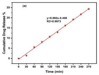
The present study investigates the kinetic and transport behavior of polyethylene Glycol–Chitosan (CS) modified Nano-Niosomal carriers for controlled Galantamine Hydrobromide (GH) delivery in Alzheimer’s Disease (AD) therapy. Polyethylene Glycolated (PEGylated) CS-coated Nano-Niosomes were successfully fabricated using Span 60 and Cholesterol through the thin-film hydration method followed by sonication. The developed nano formulation was physicochemically characterized in terms of particle size, Polydispersity Index (PDI), zeta potential, Encapsulation Efficiency (EE), permeability stability, in vitro drug release behavior, and release Kinetics. The optimized PEG-CS-GH-NIO formulation exhibited a nanoscale particle size of approximately 111.6 nm with a low PDI value of 0.11, confirming the formation of uniformly distributed Nano-Niosomal carriers. The formulation demonstrated a high EE of 98.73% and low permeability during refrigerated storage, indicating enhanced vesicular integrity and effective drug retention. In vitro release studies revealed a biphasic release profile consisting of an initial burst release phase followed by a sustained release phase over 270 min. Mathematical modeling using zero-order, first-order, Higuchi, and Korsmeyer–Peppas equations showed that the zero-order model provided the best correlation coefficient (R² = 0.9973), indicating a nearly concentration-independent controlled release mechanism. Furthermore, the release exponent obtained from the Korsmeyer–Peppas model indicated super-case II transport behavior, suggesting that polymer relaxation, swelling, and structural rearrangement of the PEG-CS network significantly contributed to galantamine release. Overall, the findings demonstrate that PEGylated CS nano-niosomes represent a promising nanocarrier platform for sustained galantamine delivery and may provide potential advantages for future AD therapeutic strategies.
Keywords:NiosomeGalantamine hydrobromideDrug release kineticsPEGylationGlycol–ChitosanControlled release
1. Introduction
Alzheimer’s Disease (AD) is one of the most common progressive neurodegenerative disorders worldwide and represents a major public health challenge owing to the continuous increase in the aging population. The disease is characterized by progressive cognitive impairment, memory loss, and behavioral dysfunction resulting from neuronal degeneration and synaptic failure. Despite extensive research efforts, currently available therapeutic strategies primarily provide symptomatic relief rather than disease modification, and no definitive cure has yet been established. Galantamine Hydrobromide (GH), an acetylcholinesterase inhibitor approved for the treatment of mild-to-moderate AD, enhances cholinergic neurotransmission and temporarily improves cognitive function. However, its clinical effectiveness remains limited by rapid systemic clearance, relatively short biological half-life, poor stability, and fluctuations in plasma drug concentration, necessitating repeated administration and reducing patient compliance [1–5].
Recent advances in nanotechnology have provided innovative opportunities for overcoming many of the limitations associated with conventional pharmaceutical formulations. Owing to their nanoscale dimensions, high surface-area-to-volume ratio, tunable physicochemical properties, and versatile surface functionality, nanomaterials have become attractive platforms for improving drug delivery efficiency. Nanotechnology-based drug delivery systems can enhance drug stability, prolong circulation time, improve EE, provide controlled and sustained drug release, and facilitate targeted delivery while minimizing systemic side effects. These unique characteristics have positioned nanotechnology as one of the most rapidly advancing approaches for the development of next-generation therapeutic delivery systems for neurological disorders and other chronic diseases [6–9]. Among various nanocarrier platforms, lipid-based and vesicular nanosystems have attracted considerable attention because of their excellent biocompatibility and structural flexibility. In particular, niosomes are nonionic surfactant-based nanovesicles composed primarily of nonionic surfactants and Cholesterol, capable of encapsulating both hydrophilic and hydrophobic therapeutic agents. Compared with conventional liposomes, niosomes exhibit superior chemical stability, lower production costs, reduced toxicity, and enhanced storage stability, making them highly promising candidates for controlled drug delivery applications. Furthermore, the nanoscale size of niosomes facilitates improved interaction with biological membranes and contributes to enhanced drug transport and sustained therapeutic performance [10–13].
Surface engineering has become an effective strategy for further improving the physicochemical properties and biological performance of nanoscale drug carriers. Polyethylene Glycol (PEG) is widely employed to modify nanoparticle surfaces because PEGylation introduces steric stabilization, minimizes particle aggregation, reduces protein adsorption, and prolongs systemic circulation by decreasing recognition by the reticuloendothelial system. In parallel, Chitosan (CS), a naturally occurring cationic polysaccharide, has attracted significant interest due to its excellent biocompatibility, biodegradability, mucoadhesive properties, and ability to regulate drug diffusion. The combination of PEG and CS creates a multifunctional polymeric coating capable of simultaneously improving vesicle stability, structural integrity, and controlled drug release characteristics [14–17]. The overall design concept of the PEG-CS modified Nano-Niosomal platform and the experimental workflow adopted in this study are schematically illustrated in Fig. 1.
Although numerous investigations have reported the fabrication of Polyethylene Glycolated (PEGylated) vesicles or CS-coated niosomes, relatively few studies have systematically examined the transport mechanisms governing drug release from PEGylated CS-modified niosomal nanocarriers. A detailed understanding of release kinetics is essential for optimizing nanocarrier performance, predicting drug transport behavior, and designing efficient sustained-release systems. Mathematical kinetic models provide valuable insight into diffusion mechanisms, polymer relaxation, matrix erosion, and other physicochemical processes that collectively determine the release profile of encapsulated therapeutics.
Therefore, the present study focuses on the preparation and comprehensive kinetic evaluation of PEGylated CS-coated niosomal nanocarriers loaded with GH. The prepared nanoformulation was investigated in terms of EE, permeability, stability, in vitro release behavior, and transport Kinetics. Furthermore, zero-order, first-order, Higuchi, and Korsmeyer–Peppas mathematical models were employed to elucidate the underlying release mechanism and to assess the suitability of PEGylated CS niosomes as sustained nanocarriers for AD therapy.
This figure is shown in the PDF version of the article.
2. Materials and Methods
2.1 Materials
GH, Span 60, Cholesterol, PEG, molecular weight 2000 Da, CS, ethanol, acetic acid, and Phosphate-Buffered Saline (PBS) (pH 7.4) were used in this study. All chemicals were of analytical grade and used without further purification. GH, Span 60, Cholesterol, PEG molecular weight 2000 Da, CS, ethanol, acetic acid, and PBS (pH 7.4) were used in this study. All chemicals were of analytical grade and used without further purification. Deionized water was used throughout all experimental procedures. All experiments were performed in triplicate to ensure the reproducibility and reliability of the obtained results.
2.2 Preparation of Polyethylene Glycolated Niosomes
Niosomal carriers containing GH were prepared using the thin-film hydration method [16], [17]. Span 60 and Cholesterol were dissolved in ethanol under continuous stirring. PEG was added to the mixture, followed by galantamine incorporation. The solvent was evaporated under reduced pressure at 40°C to form a thin lipid film. Hydration was carried out using PBS (pH 7.4). The obtained suspension was sonicated for 10 min at 40 kHz to produce nanosized vesicles.
PEGylated nano-niosomes containing GH (PEG-GH-NIO) were prepared using the thin-film hydration method [16], [17]. Span 60 and Cholesterol were dissolved in ethanol under continuous stirring. PEG was subsequently added to the lipid mixture, followed by the incorporation of GH. The organic solvent was evaporated under reduced pressure at 40°C to produce a thin lipid film on the inner wall of the flask. Hydration was carried out using PBS (pH 7.4), and the resulting suspension was sonicated for 10 min at 40 kHz to produce uniformly dispersed nanosized vesicles.
The present study investigates the kinetic and transport behavior of PEG–CS modified nano-niosomal carriers for controlled GH delivery in AD therapy. suspension was allowed to equilibrate at room temperature prior to subsequent surface modification and physicochemical characterization.
2.3 Chitosan Surface Modification
PEGylated niosomes were coated using a CS solution prepared in dilute acetic acid. The suspension was stirred continuously for 4 h at room temperature to obtain PEG-CS-GH-NIO formulations. The electrostatic interaction between the positively charged amino groups of CS and the PEGylated niosomal surface generated a stable polymeric coating surrounding the nanovesicles. This surface modification was expected to improve colloidal stability, reduce premature drug leakage, and provide a sustained drug release profile.
Following preparation, the PEG-CS-GH-NIO nanoformulations were subjected to comprehensive physicochemical characterization prior to EE and release studies.
2.4 Physicochemical Nanocharacterization
The physicochemical properties of the prepared PEG-CS-GH-NIO nanoformulations were characterized in terms of particle size, Polydispersity Index (PDI), and zeta potential. These analyses were performed to evaluate the nanoscale characteristics, particle size distribution, and colloidal stability of the developed nanoformulations prior to encapsulation and drug release studies.
2.4.1 Particle size, polydispersity index, and zeta potential analysis
The average hydrodynamic particle size, PDI, and zeta potential of PEG-CS-GH-NIO nanoformulations were determined using Dynamic Light Scattering (DLS) with a Zetasizer Nano ZS instrument (Malvern Panalytical, UK) at 25°C. Prior to measurement, samples were appropriately diluted with deionized water to minimize multiple light scattering effects. Each measurement was performed in triplicate, and the results were expressed as mean ± standard deviation.
Particle size measurements were used to determine the hydrodynamic diameter of the nano-niosomes, whereas the PDI values were employed to evaluate the uniformity of particle size distribution. Zeta potential measurements were conducted to assess the colloidal stability of the nano formulations, where higher absolute zeta potential values indicated greater electrostatic stability and lower aggregation tendency.
2.5 Encapsulation Efficiency
Encapsulation Efficiency (EE%) was determined spectrophotometrically using UV-visible analysis. The percentage of encapsulated galantamine was calculated according to Eq. (1):
C1 − C2
EE = × 100, (1)
where C₁ and C₂ represent the total and free drug concentrations, respectively [18]. EE measurements were performed after completion of physicochemical characterization to ensure that the prepared nano formulations possessed suitable nanoscale properties and colloidal stability before drug loading evaluation.
2.6 Drug Permeability Study
The stability of the prepared PEG-CS-GH-NIO nano formulations during refrigerated storage at 4°C was investigated. The permeability percentage was calculated from changes in EE over time.
Briefly, the nano formulations were stored at 4°C, and samples were collected at predetermined time intervals to evaluate drug leakage from the nanovesicles. The permeability percentage was calculated based on the reduction in EE relative to the initial value. Low permeability values were considered indicative of enhanced membrane integrity and improved physicochemical stability of the PEGylated CS-coated nano-niosomes during storage.
All measurements were performed in triplicate, and the results were reported as mean ± standard deviation.
2.7 In Vitro Drug Release
Drug release experiments were performed using the dialysis membrane technique in PBS (pH 7.4) at 37 ± 1°C under continuous shaking.
Briefly, an appropriate volume of PEG-CS-GH-NIO nano formulation containing a known amount of GH was transferred into a dialysis membrane (molecular weight cut-off: 12–14 kDa). The sealed membrane was immersed in PBS maintained at physiological temperature (37 ± 1°C) under continuous magnetic stirring to simulate physiological release conditions. Samples were withdrawn at predetermined intervals up to 270 min and analyzed spectrophotometrically [19–22]. An equal volume of fresh preheated PBS was immediately replaced after each sampling to maintain sink conditions throughout the release experiment. The cumulative percentage of galantamine released from the nano-niosomes was calculated as a function of time. All release experiments were performed in triplicate, and the obtained results were expressed as mean ± standard deviation.
2.8 Kinetic Modeling
Release data were fitted using four mathematical kinetic models to investigate the mechanism governing galantamine release from the PEG-CS-GH-NIO nano formulations Eqs. (2)-(5) [20–22].
Zero-order model:
M(t)
= k0t. (2)
M(∞) First-order model:
M(t)
−k1t
= e . (3)
M(∞) Higuchi model:
M(t)
= k t2. (4)
M(∞)
Korsmeyer–Peppas model:
M(t)
= kPt . (5)
M(∞)
M(t)
where represents the cumulative fraction of drug released at time t, k0, k1, kH, and kP are the
M(∞)
corresponding kinetic constants, and n denotes the release exponent that describes the dominant transport mechanism. The goodness-of-fit of each kinetic model was evaluated using the coefficient of determination (R²). The model exhibiting the highest R² value was considered to best describe the release kinetics of galantamine from the PEG-CS-GH-NIO nano formulations.
Furthermore, the release exponent (n) obtained from the Korsmeyer–Peppas equation was used to distinguish the dominant release mechanism, including Fickian diffusion, anomalous transport, Case II transport, and Super Case II transport, thereby providing mechanistic insight into drug diffusion and polymer relaxation within the PEGylated CS Nano-Niosomal matrix.
3. Results and Discussion
3.1 Physicochemical Characterization of Polyethylene Glycol-Chitosan-Galantamine Hydrobromide Nano-Niosomes
The physicochemical properties of the prepared PEG-CS-GH nano-niosomes were evaluated by determining the particle size, PDI, and zeta potential. The obtained results are summarized in Table 1.
The developed PEG-CS-GH nano-niosomes exhibited an average hydrodynamic particle size of 111.6 nm, confirming the successful fabrication of nanosized vesicular carriers. The obtained particle size indicates that the applied thin-film hydration method followed by PEGylation and CS surface modification was effective in producing nano-niosomes within an appropriate size range for controlled drug delivery applications. Nanocarriers with nanoscale dimensions provide a high surface-area-to-volume ratio, which can enhance drug encapsulation, improve interaction with biological interfaces, and facilitate controlled transport of therapeutic molecules.
The prepared nano-niosomes showed a PDI value of 0.11, indicating a narrow particle size distribution and high dispersion homogeneity. In general, PDI values below 0.20 represent a relatively uniform nanoparticle population, suggesting that the developed PEG-CS-GH formulation possessed good size uniformity and limited aggregation. The low PDI value further confirms the effectiveness of the preparation process in generating stable and homogeneous nano-vesicular systems.
The measured zeta potential of +2.83 mV indicated a slightly positive surface charge of the PEG-CS-GH nano-niosomes. Although the electrostatic contribution to stabilization was limited, the stability of the developed nanoformulation can be attributed mainly to the steric stabilization provided by PEG chains and the protective polymeric layer formed by CS. These surface modifications may reduce particle aggregation and improve vesicle integrity, supporting the observed stability during subsequent permeability and drug release studies.
The obtained physicochemical characteristics confirmed the successful preparation of PEG-CS-GH nano-niosomes with suitable nanoscale dimensions, narrow size distribution, and appropriate physicochemical properties for sustained GH delivery.
Table 1. Physicochemical characteristics of PEG-CS-GH nano-niosomes determined by DLS analysis.
This table is shown in the PDF version of the article.
3.2 Encapsulation Efficiency
The EE of the prepared PEG-CS-GH nano-niosomes was evaluated to determine the ability of the developed nanocarrier system to entrap GH within the vesicular structure. The PEG-CS modified niosomal formulation exhibited a high EE of 98.73%, indicating effective incorporation and retention of galantamine within the niosomal bilayer.
The high EE can be attributed to the physicochemical characteristics of the niosomal system and the synergistic contribution of its structural components. Span 60, containing long saturated alkyl chains, enhances hydrophobic interactions within the vesicular membrane and contributes to improved drug accommodation and retention. Cholesterol incorporation further improves membrane rigidity and reduces excessive permeability, thereby minimizing premature drug leakage from the nanocarrier structure.
Moreover, the surface modification of niosomes with PEG and CS played an important role in improving drug entrapment efficiency. PEG chains provide steric stabilization around the Nano-Niosomal surface, reducing vesicle aggregation and maintaining structural integrity. In addition, the CS coating forms a protective polymeric layer around the vesicles, which can restrict drug diffusion from the internal aqueous core and enhance the retention of galantamine within the carrier system. The combination of nanoscale particle dimensions, uniform size distribution, and polymer-modified vesicular architecture contributed to the high encapsulation performance of the PEG-CS-GH nano-niosomes. The obtained results demonstrate the suitability of the developed nanocarrier system for efficient galantamine loading and controlled drug delivery applications.
3.3 Permeability and Storage Stability
The permeability profile of the prepared PEG-CS-GH nano-niosomes was evaluated to investigate the ability of the developed nanocarrier system to retain GH during storage conditions. The obtained results demonstrated excellent storage stability with minimal drug leakage from the Nano-Niosomal structure under refrigerated conditions. The permeability percentage increased gradually from 0.47% after 1 h to approximately 1.04% after 3 h, indicating limited diffusion of the encapsulated drug from the vesicular system (Fig. 2).
The low permeability observed in PEG-CS-GH nano-niosomes can be attributed to the protective effect of the polymer-modified vesicular membrane. The interaction between PEG chains and CS molecules likely contributed to the formation of a compact polymeric layer surrounding the niosomal surface, which enhanced membrane integrity and reduced the mobility of drug molecules across the vesicle interface. In addition, Cholesterol incorporation improved the rigidity of the lipid bilayer, further restricting premature drug leakage during storage. The combination of PEG steric stabilization and CS polymeric coating provided an additional barrier against structural disruption of the nano-niosomes. This protective effect is particularly important for maintaining drug retention before administration and ensuring predictable release behavior during subsequent in vitro release studies.
The low permeability values confirm the ability of the PEG-CS modified Nano-Niosomal system to preserve galantamine within the carrier structure and demonstrate its potential as a stable nanocarrier platform for controlled drug delivery applications.
This figure is shown in the PDF version of the article.
3.4 In Vitro Drug Release Behavior
The in vitro release profile of GH from PEG-CS-GH nano-niosomes was investigated to evaluate the ability of the developed nanocarrier system to provide controlled drug delivery. The obtained release profile demonstrated a biphasic release pattern, consisting of an initial rapid release phase followed by a slower and sustained release stage.
During the first 90 min, an initial burst release was observed, which can be attributed to the desorption of drug molecules weakly associated with the outer surface of the nano-niosomes or located near the vesicle interface. This initial phase is commonly observed in vesicular drug delivery systems and is related to the release of readily accessible drug molecules without requiring significant structural changes within the carrier. Following the initial burst phase, the release rate gradually decreased, resulting in a prolonged and sustained release behavior. This second phase can be mainly associated with the controlled diffusion of encapsulated galantamine through the PEG-CS modified vesicular bilayer. The presence of PEG and CS coatings likely increased the diffusional resistance of the Nano-Niosomal membrane by forming a protective polymeric barrier, thereby slowing drug migration from the internal vesicular compartment into the surrounding medium. At the end of the release experiment (270 min), the cumulative release of galantamine reached 24.3%, confirming the sustained release capability of the PEG-CS modified Nano-Niosomal system (Fig. 3). The relatively slow-release rate suggests that the polymer-modified nanocarrier effectively retained the encapsulated drug and prevented rapid drug diffusion, which is desirable for maintaining prolonged therapeutic availability.
The observed biphasic release behavior indicates that PEG-CS-GH nano-niosomes possess a controlled release mechanism governed by the combined effects of drug diffusion, polymeric barrier resistance, and structural characteristics of the modified vesicular system.
This figure is shown in the PDF version of the article.
Data are expressed as the mean ± SD (n = 3).
3.5 Kinetic Analysis
Mathematical kinetic models were applied to investigate the mechanism governing galantamine release from the PEG-CS-GH Nano-Niosomal system. The experimental release data were fitted using four commonly used kinetic models, including zero-order, first-order, Higuchi, and Korsmeyer–Peppas models, to identify the dominant transport mechanism involved in drug release (Fig. 4).
The zero-order kinetic model exhibited the highest correlation coefficient (R² = 0.9973) among the evaluated models, indicating that galantamine release occurred at an approximately constant rate independent of the remaining drug concentration within the Nano-Niosomal system. This release behavior is highly desirable for controlled drug delivery applications because it can provide a more consistent drug supply and minimize fluctuations in therapeutic concentration over time.
The first-order kinetic model showed lower correlation compared with the zero-order model, suggesting that concentration-dependent release was not the predominant mechanism controlling galantamine release from the PEG-CS-GH nano-niosomes. Therefore, drug dissolution or concentration gradients alone could not fully explain the observed release pattern.
The Higuchi model demonstrated relatively high correlation values, indicating that diffusion contributed significantly to the release process. However, the lower fitting performance compared with the zero-order model suggests that diffusion was not the only mechanism involved in drug transport. The presence of PEG and CS surface modification likely introduced additional resistance to drug migration through the polymer-modified vesicular structure.
The Korsmeyer–Peppas model revealed a release exponent value of n = 1.23, indicating a super-case II transport mechanism. This type of transport behavior is generally associated with polymer relaxation, swelling, and structural rearrangement of polymeric networks. In the present system, the relaxation of the PEG-CS coating and possible reorganization of the polymeric layer likely contributed to the controlled release of encapsulated galantamine in addition to diffusion effects.
The kinetic analysis confirms that the release behavior of PEG-CS-GH nano-niosomes was controlled by a combination of diffusion and polymer relaxation mechanisms. The superior fitting of the zero-order model and the super-case II transport behavior demonstrate that PEG-CS modification effectively regulated drug transport and generated a controlled nano-delivery platform for sustained galantamine release.
This figure is shown in the PDF version of the article.
4. Conclusion
In the present study, PEGylated CS-coated nano-niosomes were successfully developed as a nanoscale delivery platform for controlled release of GH. The physicochemical characterization confirmed the successful formation of PEG-CS-GH nano-niosomes with suitable nanoscale dimensions, narrow particle size distribution, and appropriate surface properties. The obtained particle size, low PDI value, and slightly positive zeta potential demonstrated the formation of a relatively uniform nano formulation with acceptable colloidal characteristics, highlighting the effectiveness of PEGylation and CS surface modification in improving the structural properties of the vesicular system.
The prepared Nano-Niosomal formulation exhibited a high EE of 98.73%, indicating efficient incorporation and retention of galantamine within the vesicular structure. The low permeability observed during refrigerated storage further confirmed the ability of the PEG-CS modified nanocarrier to preserve drug molecules and minimize premature leakage. These findings suggest that the combined effects of PEG steric stabilization, CS polymeric coating, and Cholesterol-mediated membrane rigidity contributed significantly to maintaining vesicle integrity.
The in vitro release study demonstrated a biphasic release pattern consisting of an initial burst phase followed by a prolonged sustained release phase, confirming the capability of the developed Nano-Niosomal system to regulate galantamine release over time. Kinetic analysis revealed that the release behavior was best described by the zero-order model, while the Korsmeyer–Peppas analysis indicated super-case II transport behavior. These results suggest that drug diffusion was accompanied by polymer relaxation, swelling, and structural rearrangement within the PEG-CS network, leading to a controlled transport mechanism.
The findings demonstrate that PEG-CS-modified nano-niosomes represent a promising nanocarrier platform for sustained galantamine delivery. The combination of nanoscale characteristics, high drug loading efficiency, improved storage stability, and controlled release performance highlights the potential of this system for enhancing the delivery efficiency of therapeutic agents in AD treatment. Further investigations involving biological evaluation, cellular uptake studies, and in vivo pharmacokinetic assessment are recommended to confirm the translational potential of this nano-delivery platform.
Authors' Contributions
The author was responsible for all stages of the research and manuscript preparation and approved the final version.
Funding
This research was not supported by any specific grant from funding bodies in the public, commercial, or not-for-profit sectors.
Data Availability
All data are included in the text.
Conflict of Interest
There are no competing interests to declare.
Consent for Publication
The author confirms consent for the publication of this work
Ethics Approval and Consent to Participate
This article does not contain any studies with human participants performed by the author.
Figures
References
- [1] Gadhave, D. G., Sugandhi, V. V., Jha, S. K., Nangare, S. N., Gupta, G., Singh, S. K., ... & Paudel, K. R. (2024). Neurodegenerative disorders: Mechanisms of degeneration and therapeutic approaches with their clinical relevance. Ageing research reviews, 99, 102357. https://doi.org/10.1016/j.arr.2024.102357
- [2] Ivanova, M., Panayotova-Ovcharova, L., Nedyalkova-Petkova, D., Petkov, P., Boshev, G., & Vladeva, E. (2025). Galantamine as a potential treatment for peripheral nerve injuries: Emphasis on non-systemic delivery. Future pharmacology, 5(4), 63. https://doi.org/10.3390/futurepharmacol5040063
- [3] Suh, G. H., Jung, H. Y., Lee, C. U., Oh, B. H., Bae, J. N., Jung, H. Y., ... & Korean Galantamine Study Group. (2004). A prospective, double-blind, community-controlled comparison of three doses of galantamine in the treatment of mild to moderate Alzheimer's disease in a Korean population. Clinical therapeutics, 26(10), 1608-1618. https://doi.org/10.1016/j.clinthera.2004.10.006
- [4] Singh, B., Day, C. M., Abdella, S., & Garg, S. (2024). Alzheimer’s disease current therapies, novel drug delivery systems and future directions for better disease management. Journal of controlled release, 367, 402–
- [424] https://doi.org/10.1016/j.jconrel.2024.01.047
- [5] Wang, K., Yang, R., Li, J., Wang, H., Wan, L., & He, J. (2025). Nanocarrier-based targeted drug delivery for Alzheimer’s disease: Addressing neuroinflammation and enhancing clinical translation. Frontiers in pharmacology, 16, 1591438. https://doi.org/10.3389/fphar.2025.1591438
- [6] Majumder, J., Taratula, O., & Minko, T. (2019). Nanocarrier-based systems for targeted and site specific therapeutic delivery. Advanced drug delivery reviews, 144, 57–77. https://doi.org/10.1016/j.addr.2019.07.010
- [7] Alam, M. A. (2026). Emerging smart nanocarrier based drug delivery systems for cancer therapeutics. Next nanotechnology, 9, 100387. https://doi.org/10.1016/j.nxnano.2026.100387
- [8] Kushnazarova, R., Mirgorodskaya, A., Lenina, O., Petrov, K., Lyubina, A., Voloshina, A., ... & Zakharova, L. (2025). Niosomes and tocosomes as promising nanocontainers for the antidiabetic drug repaglinide. Materials today chemistry, 50, 103223. https://doi.org/10.1016/j.mtchem.2025.103223
- [9] Kazi, K. M., Mandal, A. S., Biswas, N., Guha, A., Chatterjee, S., Behera, M., & Kuotsu, K. (2010). Niosome: A future of targeted drug delivery systems. Journal of advanced pharmaceutical technology & research, 1(4),
- [374] https://doi.org/10.4103/0110-5558.76435
- [10] Jawale, J. K., & Kashikar, V. S. (2022). Niosomes: A promising nonionic surfactant vesicular system for enhancement of anticancer effect of phytochemicals. International journal of health sciences, 6(S5), 8809–8833.
- [11] Saraswathi, T. S., Mothilal, M., & Jaganathan, M. K. (2019). Niosomes as an emerging formulation tool for drug delivery-a review. International journal of applied pharmaceutics, 11(2), 7–15. http://dx.doi.org/10.22159/ijap.2019v11i2.30534
- [12] Rangsimawong, W., Opanasopit, P., Rojanarata, T., Duangjit, S., & Ngawhirunpat, T. (2016). Skin transport of hydrophilic compound-loaded PEGylated lipid nanocarriers: Comparative study of liposomes, niosomes, and solid lipid nanoparticles. Biological and pharmaceutical bulletin, 39(8), 1254–1262. https://doi.org/10.1248/bpb.b15-00981
- [13] Lei, W., Song, J., Chen, Y., Wang, X., Yang, S., Tan, W., ... & Gao, Z. (2025). Edible polyethylene glycol modified chitosan nanoparticles: Separation and controlled release of tea polyphenols from multiphasic systems. International journal of biological macromolecules, 315, 144375. https://doi.org/10.1016/j.ijbiomac.2025.144375
- [14] Baniasad, A., Baei, M. S., & Tala-Tapeh, S. M. (2025). Chitosan-PEGylated niosomes and liposomes as biomacromolecule carriers for Alzheimer’s disease treatment: Galantamine drug delivery carrier. Materials chemistry and physics, 352, 132003. https://doi.org/10.1016/j.matchemphys.2025.132003
- [15] Cheng, R., Xu, T., Wang, C., & Gan, C. (2021). The stabilization and antioxidant performances of coenzyme Q10-loaded niosomes coated by PEG and chitosan. Journal of molecular liquids, 325, 115194. https://doi.org/10.1016/j.molliq.2020.115194
- [16] Niknejad, K., Sharifzadeh, B. M., & Motallebi, T. T. S. (2018). Synthesis of metformin hydrochloride nanoliposomes: Evaluation of physicochemical characteristics and release kinetics. International journal of nano dimension, 9(3), 298–313. (In Persian). https://www.sid.ir/paper/322408/en
- [17] Turkez, H., Oner, S., Yıldırım, O. C., Arslan, M. E., Dimmito, M. P., Kahraman, Ç. Y., ... & Mardinoglu, A. (2025). Synthesis and characterization of memantine-loaded niosomes for enhanced Alzheimer’s disease targeting. Pharmaceutics, 17(2), 267. https://doi.org/10.3390/pharmaceutics17020267
- [18] Lu, Q., Lu, P. M., Piao, J. H., Xu, X. L., Chen, J., Zhu, L., & Jiang, J. G. (2014). Preparation and physicochemical characteristics of an allicin nanoliposome and its release behavior. LWT-food science and technology, 57(2), 686-695. https://doi.org/10.1016/j.lwt.2014.01.044
- [19] Nanaki, S. G., Spyrou, K., Bekiari, C., Veneti, P., Baroud, T. N., Karouta, N., ... & Bikiaris, D. N. (2020). Hierarchical porous carbon—plla and plga hybrid nanoparticles for intranasal delivery of galantamine for Alzheimer’s disease therapy. Pharmaceutics, 12(3), 227. https://doi.org/10.3390/pharmaceutics12030227
- [20] Georgieva, D., Nikolova, D., Vassileva, E., & Kostova, B. (2023). Chitosan-based nanoparticles for targeted nasal galantamine delivery as a promising tool in Alzheimer’s disease therapy. Pharmaceutics, 15(3), 829. https://doi.org/10.3390/pharmaceutics15030829
- [21] Tapeh, S. M. T., Baei, M. S., & Keshel, S. H. (2021). Synthesis of thermogel modified with biomaterials as carrier for hUSSCs differentiation into cardiac cells: Physicomechanical and biological assessment. Materials science and engineering: C, 119, 111517. https://doi.org/10.1016/j.msec.2020.111517
- [22] Chandrasekhar, P., & Kaliyaperumal, R. (2026). Development of transfersomal buccal patches of galantamine for enhanced bioavailability in Alzheimer’s disease therapy. Journal of applied pharmaceutical research, 14(2), 61–75. https://doi.org/10.69857/joapr.v14i2.1523