
Bioactivity Assessment of Liposome-Encapsulated Alhagi Maurorum Extract: Antioxidant and Antibacterial Activities
1. Introduction
Medicinal plants have attracted considerable attention as natural sources of biologically active compounds with potential applications in pharmaceutical, nutraceutical, and biomedical fields. Unlike many single synthetic compounds, plant extracts contain complex mixtures of secondary metabolites that may act through multiple biological mechanisms. Flavonoids, phenolic compounds, alkaloids, terpenoids, tannins, and other phytochemicals have been widely investigated because of their antioxidant, antibacterial, anti-inflammatory, and other biological activities. Growing interest in plant-derived bioactive compounds has also encouraged the development of delivery systems capable of improving the physicochemical properties and biological performance of these complex natural products [1–7].
Oxidative stress is one of the important biological processes associated with cellular and tissue damage. An imbalance between the production of reactive oxygen species and the capacity of endogenous antioxidant systems may result in oxidative modification of lipids, proteins, and nucleic acids. Although reactive oxygen species are involved in normal cellular signaling and physiological processes, excessive accumulation of these species can contribute to oxidative damage and may be associated with the development or progression of several chronic disorders. Consequently, identifying natural sources with antioxidant potential has become an important area of research. Plant-derived phenolic and flavonoid compounds are particularly interesting because of their ability to interact with free radicals and participate in different antioxidant mechanisms [8– 12].
At the same time, bacterial infections remain an important challenge in biomedical and pharmaceutical research. The increasing occurrence of bacterial strains with reduced susceptibility to conventional antimicrobial agents has stimulated the search for alternative or complementary antibacterial compounds. Plant extracts have been extensively investigated in this context because their chemically diverse constituents may exert antibacterial effects through different mechanisms, including interactions with bacterial cell membranes, alteration of membrane permeability, interference with cellular metabolism, and inhibition of essential cellular processes. However, the biological activity of a crude plant extract depends strongly on its chemical composition, extraction procedure, concentration, and experimental conditions [10–16].
Among medicinal plants, species belonging to the genus Alhagi have received attention because of their diverse phytochemical composition and traditional medicinal applications. Alhagi is a genus of the Fabaceae family distributed in several regions of Asia, Europe, and other parts of the world. Different Alhagi species have been investigated for their potential antioxidant and pharmacological activities. Reported constituents of the genus include flavonoids, alkaloids, steroids, phospholipids, polysaccharides, and other secondary metabolites. Various biological properties, including antioxidant, anti-inflammatory, hepatoprotective, antispasmodic, antidiarrheal, antinociceptive, antipyretic, antibacterial, and antifungal activities, have been associated with different species and plant parts.
A. Maurorum is one of the best-known species of this genus and has been traditionally used in different medicinal applications. The presence of multiple classes of phytochemicals makes A. Maurorum a potentially valuable source of bioactive compounds. However, as with many botanical extracts, the biological performance of an A. Maurorum extract may be influenced by the solubility, stability, chemical interactions, and bioavailability of its individual constituents. In addition, some bioactive compounds present in plant extracts may be susceptible to degradation during storage or under environmental conditions such as light, temperature, and oxygen exposure. Therefore, strategies that can improve the physicochemical handling and delivery of plant-derived compounds may be useful for enhancing their practical applications [9–12].
One promising approach is the incorporation of plant extracts into lipid-based delivery systems. Liposomes are spherical vesicular structures generally composed of phospholipid bilayers surrounding an aqueous compartment. Their amphiphilic structure allows them to accommodate a broad range of compounds with different physicochemical characteristics. Hydrophilic compounds can be associated with the aqueous internal phase, whereas lipophilic compounds can interact with the lipid bilayer. This structural versatility has made liposomes widely investigated as carriers for pharmaceutical, nutraceutical, cosmetic, and other biologically active compounds [17–21].
The incorporation of a plant extract into Liposomes may provide several potential advantages compared with the use of the free extract. Encapsulation can physically associate bioactive constituents with a lipidic carrier and may influence their dispersion in aqueous media, protect susceptible compounds from environmental degradation, and facilitate their delivery to biological interfaces. However, the effectiveness of a Liposomal formulation depends on its physicochemical properties. Consequently, characterization of the resulting vesicles is an essential part of the development of a liposome-based plant extract formulation [11].
Particle size is one of the most commonly evaluated characteristics of Liposomal systems. It can influence the physical behavior, stability, biological interaction, and delivery characteristics of vesicular formulations. Dynamic Light Scattering (DLS) is widely used for routine determination of the hydrodynamic particle size of liposomes because it provides a rapid measurement of the average particle diameter and size distribution. The Polydispersity Index (PDI) provides additional information regarding the heterogeneity of the particle population. Lower PDI values generally indicate a more uniform particle-size distribution, whereas higher values suggest greater heterogeneity within the formulation [15].
Zeta potential is another important physicochemical parameter used to characterize Liposomal dispersions. It provides an indirect indication of the surface charge of particles and can be related to electrostatic interactions between vesicles. Liposomes with greater absolute surface charge may experience stronger electrostatic repulsion, which can reduce the tendency of particles to aggregate under appropriate formulation conditions. Nevertheless, zeta potential should be interpreted together with other physicochemical parameters rather than as an independent measure of formulation stability.
Encapsulation Efficiency (EE%) is particularly important for evaluating the incorporation of a plant extract into a Liposomal carrier. It represents the proportion of the initial bioactive material that becomes associated with or entrapped within the Liposomal formulation. Determination of EE% commonly involves separation of free compounds from the liposome-associated fraction followed by quantification of the relevant extract components. Centrifugation, ultrafiltration, dialysis, and chromatographic separation have all been described for this purpose [18–22].
Despite the growing interest in plant-based delivery systems, studies evaluating the biological activity of A. Maurorum after incorporation into a simple Liposomal formulation remain relatively limited compared with studies of crude plant extracts. Importantly, encapsulation does not necessarily imply improved biological activity; therefore, direct experimental comparison between the free extract and the Liposome-encapsulated extract is valuable. Such a comparison can determine whether incorporation into the lipidic carrier preserves or alters the measurable antioxidant and antibacterial properties of the plant extract [21].
Antioxidant activity can be evaluated using complementary in vitro assays because antioxidant mechanisms are not adequately represented by a single analytical method. The 2,2-Diphenyl-1-Picrylhydrazyl (DPPH) assay is a widely used radical-scavenging method based on the reduction of a stable-colored radical. The 2,2′- azinobis-(3-ethylbenzothiazoline-6-sulfonic acid) (ABTS) assay provides another radical-scavenging approach and can be applied to compounds with different polarities. The Ferric Reducing Antioxidant Power (FRAP) assay, in contrast, evaluates the reducing capacity of a sample rather than direct radical scavenging. The combination of DPPH, ABTS, and FRAP therefore provides a broader in vitro assessment of the antioxidant properties of the extract and its Liposomal formulation [17].
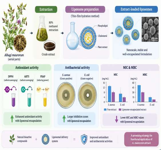
The antibacterial activity of the formulations can similarly be evaluated using complementary microbiological methods. Disk diffusion provides a simple preliminary assessment of antibacterial activity based on the inhibition zone produced around a sample-containing disk. However, diffusion-based assays are influenced by several factors, including compound diffusion through the agar matrix and the physicochemical characteristics of the tested sample. Therefore, determination of the Minimum Inhibitory Concentration (MIC) provides a quantitative estimate of the lowest concentration capable of inhibiting visible bacterial growth. The Minimum Bactericidal Concentration (MBC) can further indicate the lowest concentration associated with bactericidal activity under the defined experimental conditions. The combination of disk diffusion, MIC, and MBC therefore provides a relatively simple but informative antibacterial evaluation [17– 24]. The overall experimental concept, including A. Maurorum extraction, Liposomal encapsulation, and subsequent antioxidant and antibacterial evaluations, is schematically illustrated in Fig. 1.
The present study was designed to investigate the antioxidant and antibacterial properties of a liposome-encapsulated A. Maurorum extract and to compare its activity with that of the corresponding free extract. A simple Liposomal formulation was prepared and characterized in terms of particle size, PDI, zeta potential, and EE%. The antioxidant activity was evaluated using DPPH, ABTS, and FRAP assays, while antibacterial activity was assessed by disk diffusion, MIC, and MBC methods. The overall objective was to determine whether incorporation of A. Maurorum extract into a Liposomal carrier could provide a reproducible delivery formulation while retaining its measurable antioxidant and antibacterial activities. This approach provides a straightforward experimental framework for combining the biological potential of A. Maurorum with a simple lipid-based delivery system without requiring extensive structural or advanced nanomaterial characterization.
2. Materials and Methods
2.1 Plant Material and Preparation of Alhagi Maurorum Extract
The aerial parts of A. Maurorum were collected from Abbas Abad, Tonekabon, Mazandaran Province, Iran, in spring 2026. The plant material was washed with distilled water to remove dust and other impurities and was dried at room temperature in the shade. The dried aerial parts were ground into a fine powder using an electric laboratory grinder. For extraction, 30 g of the powdered plant material was mixed with 200 mL of 80% methanol. The mixture was kept at room temperature for 48 h with intermittent shaking. The extract was then filtered through Whatman No. 1 filter paper. The filtrate was concentrated under reduced pressure using a rotary evaporator at a temperature below 40 °C. The resulting concentrated extract was dried and stored at 4 °C in a dark container until further analysis [12–14].
The extraction yield was calculated based on the weight of the dried extract relative to the initial dry weight of the plant material.
2.2 Chemicals and Reagents
Soy phosphatidylcholine, cholesterol, methanol, ethanol, DPPH, ABTS, 2,4,6-Tripyridyl-S-Triazine (TPTZ), ferric chloride, sodium acetate, ferrous sulfate, and other analytical-grade reagents were obtained from Merck (Darmstadt, Germany). Mueller–Hinton Agar (MHA) and Mueller–Hinton Broth (MHB) were obtained from Merck (Darmstadt, Germany). All aqueous solutions were prepared using deionized water.
2.3 Preparation of Alhagi Maurorum Extract-Loaded Liposomes
Liposomes containing A. Maurorum extract were prepared using the thin-film hydration method. Soy phosphatidylcholine and cholesterol were used as the main lipid components at a phosphatidylcholine-to-cholesterol molar ratio of 7:3, a ratio commonly used for plant-extract-loaded Liposomal formulations. Briefly, soy phosphatidylcholine and cholesterol were dissolved in chloroform: methanol (2:1, v/v) in a round-bottom flask. The organic solvent was removed under reduced pressure at 40 °C using a rotary evaporator to form a uniform thin lipid film on the inner wall of the flask. The lipid film was subsequently maintained under vacuum for 2 h to remove residual solvent.
The lipid film was hydrated with an aqueous solution containing A. Maurorum extract at a final extract concentration of 10 mg/mL. Hydration was performed at 45 °C with gentle rotation for 30 min, resulting in the formation of a multilamellar Liposomal suspension.
The obtained suspension was subsequently sonicated using a probe sonicator at 40% amplitude for 5 min in pulse mode to reduce vesicle size and improve size distribution. Similar thin-film hydration followed by probe sonication has been routinely used for the preparation of plant-extract-loaded Liposomes. Blank Liposomes without plant extract were prepared using the same procedure and were used as the formulation control [25].
2.4 Determination of Particle Size and Polydispersity Index
The mean particle size and PDI of the prepared Liposomes were determined by DLS using a Zetasizer Nano ZS (Malvern Instruments Ltd., Malvern, UK).
Before measurement, the Liposomal dispersion was diluted 1:10 with deionized water to obtain an appropriate particle concentration for DLS analysis. Measurements were performed at 25 °C using disposable polystyrene cuvettes.
Three independent measurements were performed for each formulation. The results were expressed as mean±Standard Deviation (SD). Particle size was reported as hydrodynamic diameter in nanometers (nm), and PDI was reported as a dimensionless value. DLS-based measurement of particle size and PDI using a Zetasizer system is routinely employed for characterization of plant-extract-loaded Liposomes [21].
2.5 Determination of Zeta Potential
The zeta potential of the Liposomal formulations was determined using the same Zetasizer Nano ZS instrument (Malvern Instruments Ltd., Malvern, UK) by electrophoretic light scattering.
The samples were diluted 1:10 with deionized water before analysis and transferred into folded capillary cells. Measurements were performed at 25 °C. Three independent measurements were carried out for each formulation, and the results were expressed as mean±SD in millivolts (mV) [26].
2.6 Determination of Encapsulation Efficiency
The EE% of A. Maurorum extract in the Liposomes was determined by separating the non-encapsulated extract from the Liposomal fraction.
Briefly, 1 mL of the Liposomal suspension was centrifuged for 30 min at 4°C. The supernatant containing the non-encapsulated extract was collected for analysis. The total extract content was determined by disrupting an equivalent volume of the Liposomal formulation with methanol [26].
The extract concentration in the samples was determined spectrophotometrically using a calibration curve prepared from known concentrations of A. Maurorum extract. The EE% was calculated according to the following Eq. (1):
Wt − Wf
EE(%) = × 100, (1)
Wt where Wt represents the total amount of extract added during liposome preparation, and Wf represents the amount of free, non-encapsulated extract detected in the supernatant.
All measurements were performed in triplicate.
2.7|2,2-Diphenyl-1-Picrylhydrazyl Radical Scavenging Activity
The antioxidant activity of the free A. Maurorum extract and liposome-encapsulated extract was evaluated using the DPPH radical scavenging assay. Different concentrations of the samples (25-500 µg/mL) were prepared in methanol. Briefly, 1 mL of each sample was mixed with 1 mL of freshly prepared DPPH solution (0.1 mM in methanol). The mixtures were incubated in the dark at room temperature for 30 min. The absorbance was then measured at 517 nm using a UV-visible spectrophotometer [27].
Methanol and DPPH solution without sample were used as the blank and control, respectively. The percentage of DPPH radical scavenging activity was calculated according to the following Eq. (2):
Ac − As
DPPH scavenging activity (%) = × 100, (2)
Ac where AC is the absorbance of the control, and AS is the absorbance of the sample. All measurements were performed in triplicate.
2.8|2,2′-Azinobis-(3-Ethylbenzothiazoline-6-Sulfonic Acid) Radical
Scavenging Activity
The ABTS radical scavenging activity of the free and Liposome-encapsulated A. Maurorum extracts was determined using a standard spectrophotometric method. The ABTS radical cation was prepared by mixing 7 mM ABTS solution with 2.45 mM potassium persulfate and keeping the mixture in the dark for 12–16 h at room temperature.
The resulting ABTS solution was diluted with methanol to obtain an absorbance of 0.70±0.02 at 734 nm. Then, 100 µL of each sample at different concentrations (25-500 µg/mL) was mixed with 3.9 mL of the diluted ABTS solution. After incubation for 6 min at room temperature, the absorbance was measured at 734 nm [21].
The percentage of ABTS radical scavenging activity was calculated using the following Eq. (3):
Ac − As
ABTS scavenging activity (%) = × 100, (3)
Ac where Ac represents the absorbance of the control and As represents the absorbance of the sample. All measurements were performed in triplicate.
2.9 Ferric Reducing Antioxidant Power Assay
The FRAP of the free and Liposome-encapsulated A. Maurorum extracts was determined using the FRAP assay. The FRAP reagent was freshly prepared by mixing 300 mM acetate buffer (pH 3.6), 10 mM TPTZ solution, and 20 mM ferric chloride solution at a ratio of 10:1:1 (v/v/v). Briefly, 100 µL of each sample was mixed with 3 mL of freshly prepared FRAP reagent and incubated at 37 °C for 10 min. The absorbance was measured at 593 nm using a UV–visible spectrophotometer. Ferrous sulfate was used to prepare the calibration curve, and the antioxidant capacity was expressed as µmol Fe²⁺ equivalents per gram of extract. The FRAP value was calculated based on the calibration curve obtained using ferrous sulfate standards. All measurements were performed in triplicate [28].
2.10 Bacterial Strains
The antibacterial activity of the free and Liposome-encapsulated A. Maurorum extracts was evaluated against Staphylococcus Aureus ATCC 25923 and Escherichia Coli ATCC 25922. The bacterial strains were cultured in MHB and maintained under standard microbiological conditions.
2.11 Disk Diffusion Assay
The antibacterial activity was evaluated using the disk diffusion method. Bacterial suspensions were adjusted to 0.5 McFarland turbidity and uniformly spread on MHA plates using sterile cotton swabs. Sterile paper disks were loaded with 20 µL of the free A. Maurorum extract or Liposome-encapsulated extract at a concentration of 10 mg/mL and placed on the inoculated agar plates. Blank liposomes and the corresponding solvent were used as negative controls, while gentamicin was used as the positive control. The plates were incubated at 37°C for 24 h. The diameters of the inhibition zones were measured in millimeters. All experiments were performed in triplicate [19].
2.12 Determination of Minimum Inhibitory Concentration
The MIC was determined using the broth microdilution method in 96-well microplates. Two-fold serial dilutions of the free and liposome-encapsulated extracts were prepared in Mueller–Hinton broth at concentrations ranging from 0.125 to 8 mg/mL. Each well was inoculated with a standardized bacterial suspension at approximately 5×10⁵ CFU/mL. The plates were incubated at 37°C for 24 h.
The MIC was defined as the lowest concentration of the tested sample that showed no visible bacterial growth. Growth and sterility controls were included in each experiment [21].
2.13 Determination of Minimum Bactericidal Concentration
The MBC was determined from the wells showing no visible bacterial growth in the MIC assay. A small aliquot from each clear well was transferred onto MHA plates and incubated at 37 °C for 24 h. The MBC was defined as the lowest concentration at which no visible bacterial colonies were observed after incubation. All measurements were performed in triplicate [18].
3. Results and Discussion
3.1 Characterization of Alhagi Maurorum Extract-Loaded Liposomes
The physicochemical characteristics of the A. Maurorum extract-loaded Liposomes were evaluated in terms of particle size, PDI, zeta potential, and EE%. The formulation showed nanoscale particle formation with a relatively narrow particle-size distribution, a moderately negative surface charge, and efficient extract encapsulation. The corresponding physicochemical characteristics are summarized in Table 1.
Table 1. Physicochemical characteristics of A.
This table is shown in the PDF version of the article.
The mean particle size was 160±4.8 nm, confirming the formation of nanosized Liposomal vesicles. The PDI value of 0.230±0.015 indicated a relatively homogeneous particle-size distribution. The zeta potential was - 28.6±1.3 mV, demonstrating a moderately negative surface charge that can contribute to colloidal stability. The EE% reached 82.4±2.6%, indicating effective incorporation of the A. Maurorum extract into the Liposomal formulation. Overall, the physicochemical properties presented in Table 1 demonstrated successful preparation of the extract-loaded Liposomal system.
3.2 Antioxidant Activity
The antioxidant activity of the free A. Maurorum extract and the extract-loaded Liposomes was assessed using DPPH, ABTS, and FRAP assays. The free extract exhibited measurable radical-scavenging and reducing activities, while incorporation into the Liposomal system resulted in enhanced antioxidant responses. The results obtained from the three antioxidant assays are presented in Fig. 2.
This figure is shown in the PDF version of the article.
The free A. Maurorum extract showed a DPPH IC₅₀ of 45.22±2.11 µg/mL, an ABTS radical-scavenging activity of 68.40±1.85%, and a FRAP value of 139.90±4.01 µM Fe²⁺ equivalents/g. As shown in Table 2, the extract-loaded liposomes exhibited stronger antioxidant responses than the free extract. The DPPH IC₅₀ decreased to 34.16±1.74 µg/mL following Liposomal encapsulation, while ABTS inhibition increased to 76.85±1.62% and FRAP activity increased to 165.72±4.36 µM Fe²⁺ equivalents/g. In contrast, blank Liposomes exhibited minimal antioxidant activity, with an ABTS inhibition of 3.21±0.42% and a FRAP value of 8.14±0.76 µM Fe²⁺ equivalents/g. These results indicate that the antioxidant activity of the Liposomal formulation was predominantly attributable to the encapsulated A. Maurorum extract.
3.3 Antibacterial Activity by Disk Diffusion
The antibacterial activity of the free A. Maurorum extract and extract-loaded Liposomes was evaluated against S. Aureus and E. coli using the disk diffusion method. Both formulations produced measurable inhibition zones against the tested bacterial strains, with a more pronounced inhibitory effect observed for the extract-loaded Liposomes. The antibacterial activity and inhibition-zone diameters are presented in Table 2.
Table 2. Antibacterial activity of free and Liposome-encapsulated A. Maurorum extract against S. Aureus and E. Coli.
This table is shown in the PDF version of the article.
The free A. Maurorum extract produced inhibition zones of 13.0±0.5 mm against S. Aureus and 7.0± 0.8 mm against E. Coli. As shown in Table 2, incorporation of the extract into the Liposomal system increased the inhibition-zone diameter to 26.0±0.1 mm for S. Aureus and 18.0±0.3 mm for E. Coli. The larger inhibition zones observed with the extract-loaded Liposomes indicate enhanced antibacterial activity following incorporation of the plant extract into the lipid-based delivery system. Blank Liposomes showed no detectable inhibition zone, confirming that the observed antibacterial effect was associated primarily with the encapsulated extract. The antibacterial response was more pronounced against S. Aureus than E. Coli under the tested conditions.
3.4 Minimum Inhibitory and Minimum Bactericidal Concentrations
The MIC and MBC of the free A. Maurorum extract and extract-loaded Liposomes were determined against S. Aureus and E. coli using the broth dilution method. The MIC was defined as the lowest concentration showing no visible bacterial growth, whereas the MBC was defined as the lowest concentration producing no detectable colony growth after subculturing. The resulting MIC and MBC values are summarized in Table 3.
Table 3. MIC and MBC of free and Liposome-encapsulated A. Maurorum extract.
This table is shown in the PDF version of the article.
The free extract exhibited MIC values of 12.50 mg/mL and 3.125 mg/mL against S. Aureus and E. coli, respectively. The corresponding MBC values were 50.00 mg/mL for S. Aureus and 6.50 mg/mL for E. Coli. As presented in Table 3, encapsulation of the extract into Liposomes reduced both the MIC and MBC values. For the extract-loaded Liposomes, the MIC decreased to 6.25 mg/mL against S. Aureus and 1.75 mg/mL against E. coli, while the corresponding MBC values were 25.00 mg/mL and 3.25 mg/mL, respectively. These results demonstrate that the liposome-encapsulated extract exhibited greater antibacterial potency than the free extract, as indicated by the lower concentrations required to inhibit and eliminate bacterial growth. 3.5|Comparative Evaluation of Free and Liposome-Encapsulated A.
Maurorum Extract
The overall performance of the free and Liposome-encapsulated A. Maurorum extract was compared based on antioxidant and antibacterial parameters. The Liposomal formulation showed an improved antioxidant response together with enhanced antibacterial activity against both tested bacterial strains. The comparative changes in the main biological parameters are presented in Table 5.
This figure is shown in the PDF version of the article.
The results shown in Table 5 demonstrate that Liposomal encapsulation improved the antioxidant performance of the A. Maurorum extract across all three evaluated assays. The DPPH IC₅₀ decreased from 45.22±2.11 to 34.16±1.74 µg/mL, corresponding to a 24.46% reduction. Similarly, ABTS radical-scavenging activity increased from 68.40±1.85% to 76.85±1.62%, while FRAP activity increased from 139.90±4.01 to 165.72±4.36 µM Fe²⁺ equivalents/g, representing increases of 12.35% and 18.46%, respectively.
A more pronounced improvement was observed in the antibacterial activity. Against S. aureus, the inhibition-zone diameter increased from 13.0±0.5 to 26.0±0.1 mm, corresponding to a 100% increase. Against E. coli, the inhibition zone increased from 7.0±0.8 to 18.0±0.3 mm, representing a 157.14% increase. The MIC values were reduced from 12.50 to 6.25 mg/mL for S. Aureus and from 3.125 to 1.75 mg/mL for E. Coli. The corresponding MBC values decreased from 50.00 to 25.00 mg/mL and from 6.50 to 3.25 mg/mL, respectively.
The liposome-encapsulated formulation exhibited greater biological activity than the free extract, with the most pronounced relative improvement observed in the antibacterial response. The reduction in MIC and MBC values, together with the enlargement of inhibition zones, indicates that incorporation of the extract into the Liposomal carrier enhanced its effective antibacterial performance under the tested conditions.
4. Discussion
The present study evaluated the antioxidant and antibacterial activities of A. Maurorum extract before and after incorporation into a Liposomal delivery system. The physicochemical characterization confirmed the formation of nanoscale extract-loaded Liposomes with a relatively narrow particle-size distribution, moderate surface charge, and satisfactory EE%. Importantly, Liposomal incorporation was associated with improved antioxidant and antibacterial responses compared with the free extract, suggesting that the Liposomal formulation can enhance the functional performance of bioactive compounds present in A. Maurorum extract [11–15].
The particle size and PDI values indicated the formation of a relatively homogeneous nanoscale formulation. The incorporation of the plant extract into phospholipid-based vesicles may facilitate its dispersion in the aqueous environment and improve the availability of poorly water-soluble bioactive constituents. The negative zeta potential observed for the formulation may also contribute to electrostatic repulsion between vesicles and thereby reduce aggregation during storage. The relatively high EE% further indicates that the selected phosphatidylcholine-to-cholesterol composition and thin-film hydration procedure were suitable for incorporating the extract into the Liposomal structure [12].
The antioxidant assays demonstrated enhanced activity following Liposomal encapsulation. The lower DPPH IC₅₀ value observed for the Liposome-encapsulated extract indicates greater radical-scavenging efficiency, whereas the increased ABTS and FRAP responses indicate improved overall antioxidant performance. This enhancement may be related to improved dispersion and accessibility of extract-derived phenolic and flavonoid compounds in the assay medium. In addition, the phospholipid bilayer may provide a protective microenvironment for sensitive antioxidant constituents and facilitate their gradual interaction with reactive species. The consistent trend observed across DPPH, ABTS, and FRAP assays supports the contribution of multiple antioxidant mechanisms rather than dependence on a single assay response [12–15].
The antibacterial findings also demonstrated a stronger inhibitory effect after Liposomal incorporation. The larger inhibition zones observed against both S. Aureus and E. coli indicate improved antibacterial activity of the encapsulated formulation. The reduction in MIC and MBC values further supports this observation, demonstrating that lower concentrations of the liposome-associated extract were required to inhibit and eliminate the tested bacterial strains. Enhanced antibacterial activity may result from improved dispersion of the extract, increased interaction of the Liposomal vesicles with bacterial cell surfaces, and more effective delivery of bioactive constituents to the bacterial environment [16].
The differences observed between the two bacterial strains may also reflect variations in their cell-envelope structures. S. Aureus, a Gram-positive bacterium, lacks the outer membrane characteristic of Gram-negative bacteria, whereas E. coli possesses an additional outer membrane that can act as a permeability barrier. Nevertheless, the Liposome-encapsulated extract increased the inhibition-zone diameter against both organisms, indicating that the formulation retained antibacterial activity across different bacterial envelope architectures [17].
Overall, the combined physicochemical, antioxidant, and antibacterial findings indicate that Liposomal incorporation can improve the functional performance of A. Maurorum extract. The enhanced activity observed with the Liposomal formulation, together with its nanoscale characteristics and effective extract encapsulation, supports the potential of phospholipid-based Liposomes as a delivery platform for plant-derived bioactive compounds. Further investigations involving storage stability, release kinetics, cytocompatibility, and identification of the major encapsulated phytochemicals would provide additional information regarding the applicability of this formulation.
Authors' Contributions
All aspects of the research and manuscript preparation were carried out by the author. The author has read and approved the final version of the manuscript.
Data Availability
All data supporting the reported findings in this research paper are provided within the manuscript.
Funding
Not applicable.
Conflict of Interest
The author declares that they do not have any conflict of interest.
Consent for Publication
The author confirms consent for the publication of this work.
Ethics Approval and Consent to Participate
This article does not contain any studies with human participants performed by the author.
References
- [1] Miruka, C. O. (2026). Beyond the pill: Unlocking the antidiabetic secrets of medicinal plants. https://doi.org/10.13140/RG.2.2.26465.77928
- [2] Sergeev, A. (2015). Travel photography and digital pictures: 15/1564-Shoot of camelthorn plant (Alhagi Maurorum) with small leaves in a silty depression in Al Nuaman, near Zubara. Northern Qatar. https://www.asergeev.com/pictures/archives/compress/2015/1564/15l.htm
- [3] Sergeev, A. (2015). Travel photography and digital pictures: 12/1564-Camelthorn plants (Alhagi Maurorum) in a silty depression in Al Nuaman, near Zubara. Northern Qatar. https://www.asergeev.com/pictures/archives/compress/2015/1564/12.htm
- [4] Sergeev, A. (2015). Travel photography and digital pictures: 21/1608-Flower and fruits of camelthorn (Alhagi Maurorum) taken from Rawdat Al Faras Research Station (RAFRS) near Al Zubara Road. Qatar. https://www.asergeev.com/pictures/archives/compress/2015/1608/21l.htm
- [5] Abbas, F., Iqbal, M., Khalil, O., & Rizwan, M. (2026). Green synthesis of silver nanoparticles using Alhagi Maurorum leaf extract: Characterization, antibacterial, and antioxidant activities. Planta animalia, 5(3), 19–30. https://doi.org/10.71454/PA.005.03.0481
- [6] Alotaibi, M. O., Akram, B., Umar, A., Nadeem, A., Gubanova, L., Igwegbe, C. A., ... & Ghosh, S. (2026). Alhagi Maurorum-based magnetic silver nanocatalyst synthesis and its function in the catalytic breakdown of malachite green. Applied water science, 16(4), 109. https://doi.org/10.1007/s13201-026-02770-9
- [7] Kowsarkhizi, A. S., Attar, M. A., Mafinezhad, M. S., Keshmiri, S. F., Mohammadi, F., Mostafavi, S. Z., ... & Karimi, E. (2026). Phenolic microencapsulation of Alhagi Maurorum: A novel approach to enhancing Diabetes nutrition therapy. Applied biological chemistry, 69(1), 2. https://doi.org/10.1186/s13765-025-01070-1
- [8] Mahde, M. H., & Hammod, A. J. (2024). Effect of addition aqueous extract of camel thorn (Alhagi Maurorum) plant roots and some of immune enhancers with drinking water on productive performance of broilers. IOP conference series: Earth and environmental science (Vol. 1371, No. 7, p. 072008). IOP Publishing. https://doi.org/10.1088/1755-1315/1371/7/072008
- [9] Nithikadphanich, C., & Unpaprom, Y. (2026). From orchid by-product to functional ingredient: Preliminary biosafety evaluation of pseudobulb extracts using the Allium cepa model. https://www.researchgate.net/publication/414383541_From_Orchid_By-product_to_Functional_Ingredient_Preliminary_Biosafety_Evaluation_of_Pseudobulb_Extracts_Using_the_All ium_cepa_Model
- [10] Javed, S., Kadhem, A. A., Warsi, A. Z., AlAnsari, M. S., Jabeen, N., & Imanov, H. (2026). Green synthesis of plant-extract-mediated metal nanoparticles for renewable energy conversion and storage applications. International journal of advances in signal and image sciences, 12(3s (2026)), 909–925. https://doi.org/10.29284/8agjvy82
- [11] Vinodha, S., Yuvaraj, G., Vidhya, L., & Daniel, J. E. S. (2026). Biosynthesis of ZrO2 Bioceramic from Phyllanthus niruri plant extract and it’s reinforcement effects on okra fiber-reinforced vinyl ester composite. Polymer bulletin, 83(3), 123. https://doi.org/10.1007/s00289-025-06206-3
- [12] Shaukat, F., Khalil, A. A., Akhtar, M. N., Bilal, M., Guo, Y., Huang, J., ... & Sun, X. (2026). A comprehensive insight into optimized ultrasound-assisted extraction of bioactive compounds and antioxidant activities from dandelion (Taraxacum officinale) plant extract using response surface methodology. Ultrasonics sonochemistry, 129, 107871. https://doi.org/10.1016/j.ultsonch.2026.107871
- [13] Kanchana, V., Kistan, A., Ramesh, S., & Balaramesh, P. (2026). Green synthesis of Cu wrapped NiO@ rGO nanocomposite using Acacia nilotica plant extract: A sustainable solution for photocatalytic and antioxidant activities. Waste and biomass valorization, 17(4), 2247–2260. https://doi.org/10.1007/s12649-025-03244-w
- [14] Patel, K., Shah, M., Patel, K., & Nagani, A. (2026). Recent advances in piperazine derivatives as antibacterial agents: A comprehensive review (2020-2024). Molecular diversity, 30(3), 3009–3030. https://doi.org/10.1007/s11030-025-11311-6
- [15] Lou, S., Xing, C., Zhang, J., Yang, Q., Wang, W., Qian, Y., ... & Pan, Y. (2026). Efficient organic nanoparticles for photothermal antibacterial activity and immune regulation to promote maxillofacial wound healing and scarless repair. Biomaterials, 324, 123534. https://doi.org/10.1016/j.biomaterials.2025.123534
- [16] Wan, P., Hua, T., Zhao, X., Deng, M., Chen, L., Xiao, C., & Chen, X. (2026). Tertiary alkylamine-functionalized polyaspartamides with potent antibacterial activity. Bioactive materials, 57, 445–456. https://doi.org/10.1016/j.bioactmat.2025.11.017
- [17] Ghosh, D., Bag, S., & De, P. (2026). Enzyme-responsive polymeric materials with anticancer and antibacterial activities. Biomacromolecules, 27(1), 16–58. https://doi.org/10.1021/acs.biomac.5c01944
- [18] Abdelnasser, E., Fahim, A. M., Oyekunle, D. T., Ramadan, A. E.-M. M., & Khodair, A. I. (2026). Green synthesis of 2D azine-linked covalent organic framework with antibacterial activity correlated by molecular docking study and computational calculations. Scientific reports, 16(1), 1335. https://doi.org/10.1038/s41598-025-32900-3
- [19] Lisjak, M., Špoljarević, M., Ravlić, J., Lončarić, Z., & Galić, L. (2026). Phenolic compounds and antioxidant activity: Analytical methods and current knowledge—A review. Methods and protocols, 9(2), 60. https://doi.org/10.3390/mps9020060
- [20] Duan, Y., Li, X., Zhu, Y., Chen, J., Xue, J., Wang, H., & Chen, S. (2026). Dual-frequency ultrasonic-assisted extraction of pumpkin polysaccharides: Process optimization, structural characterization, and antioxidant activity. LWT - Food science and technology, 119220. https://doi.org/10.1016/j.lwt.2026.119220
- [21] S Jabir, M., Taha, A., & Sahib, U. (2026). Antioxidant activity of Linalool. Engineering and technology journal, 36(1), 64–67. https://doi.org/10.30684/etj.36.1B.11
- [22] Zhang, K., Wang, Z., Zhang, L., Wu, H., Liu, J., Zhang, M., ... & Liu, R. (2026). Pharmacologically inherited carbon dots from Salvia miltiorrhiza with potent antioxidant activity and multi-pathway modulation for myocardial ischemia-reperfusion injury therapy. Theranostics, 16(4), 1855. https://doi.org/10.7150/thno.123141
- [23] Rane, B. R., Chaure, P. P., Bhutekar, S. K., Gawade, S. S., Mutkule, D. S., Udmale, A. V, & Jain, A. S. (2026). Recent advancements in revolutionizing cancer therapy with Liposomal drug delivery. Current cancer therapy reviews, 22(4), 392–405. https://doi.org/10.2174/0115733947379210250530103826
- [24] Krawczyńska, K., Rusak, A., Górnicki, T., Mrozowska, M., Dzięgiel, P., & Gubernator, J. (2026). Development of new Liposomal formulations of quercetin–in vitro study. Scientific reports, 16(1), 17953. https://doi.org/10.1038/s41598-026-48068-3
- [25] Syaputri, F. N., Wilar, G., Zulhendri, F., Mohammed, A. F. A., Elamin, K. M., Osman, W. J., Firmansyah, F., & Wathoni, N. (2026). Liposome-based drug delivery for Diabetes: Therapeutic applications and a nanomedicine perspective on diabetic complications. International journal of nanomedicine, 21, 1–32. https://doi.org/10.2147/IJN.S593144
- [26] Chen, J., Dehabadi, L., Vieira, A., Ma, Y. C., & Wilson, L. (2026). Next-generation nutrient delivery: Physicochemical insights into multicomponent Liposomes. International journal of technology, 17(1), 282. doi. 10.14716/ijtech.v17i1.8049
- [27] Baniasad, A., Baei, M. S., & Tala-Tapeh, S. M. (2025). Chitosan-PEGylated niosomes and liposomes as biomacromolecule carriers for Alzheimer’s disease treatment: Galantamine drug delivery carrier. Materials chemistry and physics, 352, 132003. https://doi.org/10.1016/j.matchemphys.2025.132003
- [28] Niknejad, K., Sharifzadeh, B. M., & Motallebi, T. T. S. (2018). Synthesis of Metformin Hydrochloride Nanoliposomes: Evaluation of physicochemical characteristics and release kinetics. International journal of nano dimension, 9(3), 298-313. (In Persian). https://www.sid.ir/paper/322408/en